


Scientists at the University of California, Riverside have achieved a significant milestone in bioengineering by developing the first fully synthetic model of human brain tissue. This breakthrough, named the Bijel-Integrated PORous Engineered System (BIPORES), eliminates the need for animal-derived materials and biological coatings, potentially reducing reliance on animal testing in research.
Neural tissue engineering seeks to replicate the brain’s intricate environment, known as the extracellular matrix. This matrix supports the growth and connectivity of nerve cells through its carefully structured design and signaling capabilities. While existing 3D tissue-engineered models show promise in mimicking brain functions, they often fall short in replicating the brain’s subtle design features, which are crucial for accurate cell behaviors.
The BIPORES innovation represents a leap forward. With its foundation primarily consisting of polyethylene glycol (PEG), a chemically neutral polymer, the new model provides a stable platform for cell growth. On its own, PEG is not conducive to cell attachment; it typically requires proteins such as laminin or fibrin to support cell adhesion.
To overcome limitations in previous methods that produced materials no thicker than 200 micrometers, the researchers developed a novel approach. They combined fibrous structures with intricate pore patterns inspired by bijels, which are soft materials characterized by smooth, saddle-shaped internal surfaces. This system not only enhances the physical structure of the model but also facilitates nutrient and waste exchange, crucial for supporting cell growth.
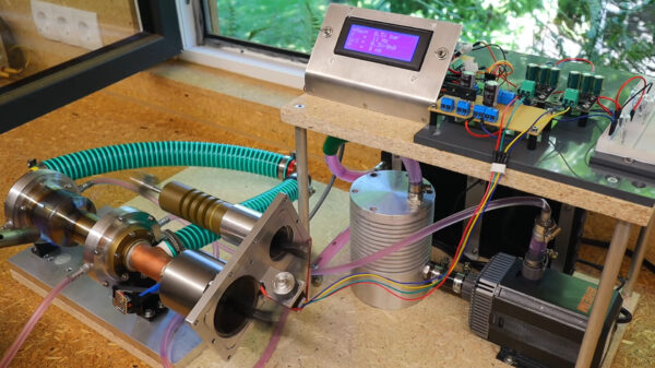
The research team utilized a specialized microfluidic setup and bioprinter to create layered, interconnected porous structures. These innovations allowed for the successful testing of neural stem cells within the new material, leading to strong cell attachment and even the formation of active nerve connections.
According to Prince David Okoro, the study’s lead author, “Since the engineered scaffold is stable, it permits longer-term studies.” This stability is vital for researching diseases and trauma, as it allows mature brain cells to better reflect actual tissue functions.
The fabrication of the scaffold involved a unique liquid mixture of PEG, ethanol, and water. The interaction of these components triggered a separation process that formed a sponge-like structure rich in pores, enabling efficient nutrient transfer. Iman Noshadi, an associate professor of bioengineering at UCR, emphasized that “the material ensures cells get what they need to grow, organize, and communicate with each other in brain-like clusters.”
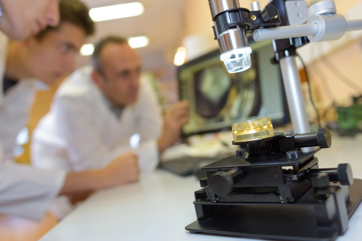
Currently, the BIPORES scaffold measures just two millimeters across, but the research team is already looking to scale it up. They have submitted further research exploring the application of this technology to other tissues, including liver tissue. The broader vision includes creating a network of lab-grown mini-organs that can interact as a cohesive system, similar to the human body’s functioning organs.
Such interconnected models could revolutionize the understanding of how different tissues respond to treatments and influence one another. Noshadi noted, “It is a step toward understanding human biology and disease in a more integrated way.”
From a biomimicry perspective, this layered fabrication method offers a more accurate representation of real brain tissue behavior. As a powerful tool for studying diseases, testing new drugs, and developing future treatments for damaged neural tissue, the BIPORES model stands to significantly advance the field of biomedical research.
The findings of this groundbreaking study have been published in Advanced Functional Materials.