


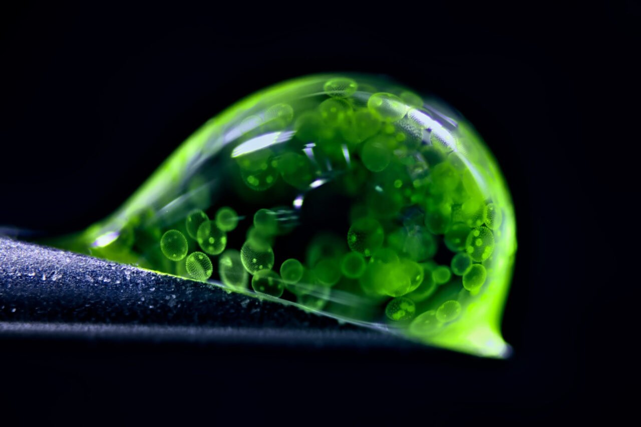
A remarkable collection of microscopic photographs has captured the imaginations of science enthusiasts and professionals alike, highlighting the intricate beauty of the unseen world. The annual Nikon Photomicrography Competition celebrated its winners, showcasing stunning images that challenge our perceptions of reality. Among the notable entries, a photograph by Jan Rosenboom, a chemical engineer from Germany, was named the runner-up for his striking depiction of colonial algae within a drop of water.
The competition invited participants from various fields to submit photographs that capture the essence of microscopy in science. Each year, the event brings together professional scientists and hobbyists, all eager to share their awe-inspiring views of familiar objects examined through an entirely new lens. Ed Cara, a judge in the 2023 competition, noted the high caliber of submissions, which ranged from cellular networks in humans to the hidden worlds within ordinary mushrooms.
Highlights from the Competition
This year’s winners featured a variety of subjects, each offering a glimpse into the microscopic realm. One standout entry came from Wim van Egmond of the Micropolitan Museum in the Netherlands, who secured ninth place with his captivating close-up of red pigments from the fungus Talaromyces purpureogenus. This fungus is a relative of the penicillin-producing Penicillium and showcases the unexpected aesthetic appeal of fungi.
Another noteworthy image was taken by researchers at the Friedrich Miescher Institute for Biomedical Research in Switzerland, which depicted a mouse colon using confocal microscopy. This technique is widely employed in biomedical research to study cells marked with fluorescent probes, illustrating the critical role that mice play in scientific exploration.
In a striking capture, James Hayes of Vanderbilt University presented heart muscle cells in the process of division. His photograph highlights the dynamic cellular networks at work within our bodies, ensuring vital functions are maintained.
One of the more surprising entries came from Stella Whittaker at the National Institutes of Health (NIH), whose image of induced pluripotent stem cell (iPSC)-derived sensory neurons resembled a swirling black hole. This dramatic representation utilized multiple microscopy techniques to convey the complexity of cellular structures.
Unexpected Visions of Nature
The competition also featured images that reveal the often-overlooked aspects of nature. One such photograph, taken by John-Oliver Dum from Germany’s Medienbunker Produktion, showcased pollen spores suspended on a spider web. Dum’s entry secured third place and exemplified the delicate yet resilient nature of life forms.
In a more somber depiction, Igor Robert Siwanowicz of the Howard Hughes Medical Institute captured the interactions of marrow pollen germinating while being parasitized by a filamentous fungus. This image illustrates the often brutal interdependencies that characterize the microscopic world.
The overall winner of this year’s competition was Zhang You, an entomologist from China, who photographed a rice weevil poised on a single grain of rice. His award-winning image, a composite of over 100 individual photographs, reflects years of dedication to ecological and insect science photography. Zhang noted that this image is the culmination of his efforts to educate others about entomology.
The Nikon Photomicrography Competition continues to inspire curiosity and appreciation for the microscopic universe, reminding us of the beauty that lies beyond our immediate perception. As technology advances, the ability to explore and document these hidden wonders will only grow, offering new insights into the natural world.